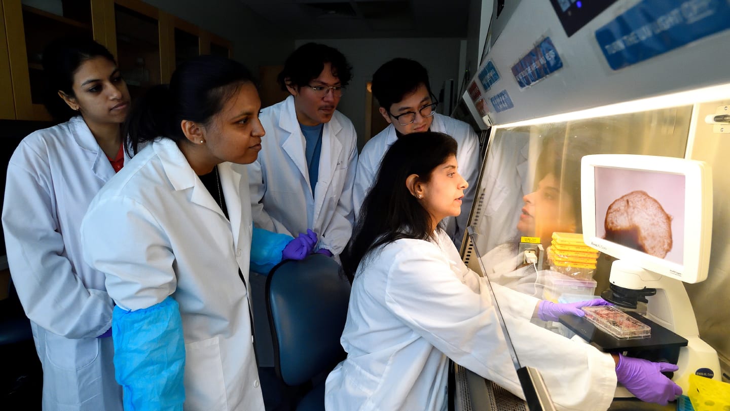
Scientists at Johns Hopkins University have grown an organoid system that integrates tissues representing multiple brain regions and vascular structures to model early human brain development.
Lead author Annie Kathuria, an assistant professor in the university’s Department of Biomedical Engineering, says: “Most brain organoids that you see in papers are one brain region, like the cortex or the hindbrain or midbrain. We've grown a rudimentary whole-brain organoid; we call it the multi-region brain organoid (MRBO).”
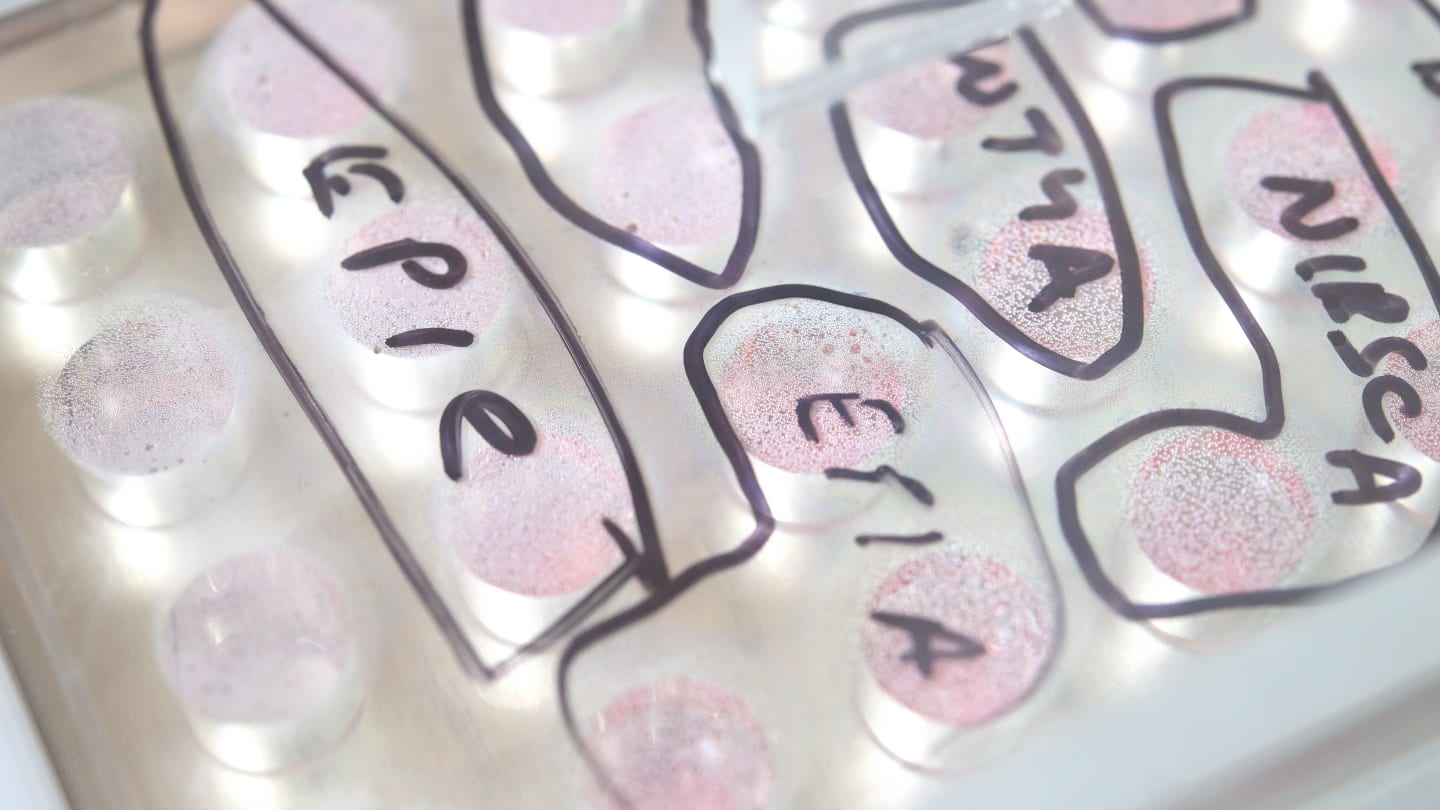
MRBO combines cerebral, midbrain, hindbrain, and endothelial components – all derived from induced pluripotent stem cells (iPSCs). It was created by first growing each regional organoid individually using protocols that guide differentiation through the manipulation of signaling pathways. For instance, mid/hindbrain organoids were exposed to BMP4 and Wnt modulators, while endothelial organoids were developed using mesoderm-inducing conditions followed by vascular growth factor supplementation. After 60 days in culture, the team found that the different regions retained their identities and formed a unified organoid that mimics early fetal brain development.
Single-nucleus RNA sequencing confirmed the presence of distinct neural and vascular cell populations from all contributing regions. A news article from HJohns Hopkins University on the work added: “The multi-region mini brain organoid retained a broad range of types of neuronal cells, with characteristics resembling a brain in a 40-day-old human fetus. Some 80% of the range of types of cells normally seen at the early stages of human brain development was equally expressed in the laboratory-crafted miniaturized brains. Much smaller compared to a real brain—weighing in at 6 million to 7 million neurons compared with tens of billions in adult brains—these organoids provide a unique platform on which to study whole-brain development.”

Rather than just using standard endothelial cells, the vascular part of the MRBO contains a range of cell types, including pericytes, angiogenic progenitors, and stromal cells, that more closely resemble real brain vasculature. These endothelial cells weren’t just along for the ride either; the study demonstrates that they play an active role in shaping brain development, particularly in the hindbrain, where they appear to support the maintenance of intermediate progenitor cells.
The researchers also measured neural activity over time and found increasing levels of network synchronization, suggesting that the different brain regions were not only integrated structurally, but also functionally. Molecular profiling and imaging backed up the findings, confirming the presence of neurons, synaptic proteins, and even early markers of blood-brain barrier development.
While the MRBOs still have limitations, such as the lack of a perfused blood supply and full connectivity between brain regions, the team sees them as a step forward for modelling neurodevelopmental disorders and testing therapies in a system that includes both neural and vascular elements. The systems could be particularly beneficial for studying diseases where vascular dysfunction intersects with neural development, such as certain forms of autism, schizophrenia, and vascular dementia.
“Diseases such as schizophrenia, autism, and Alzheimer's affect the whole brain, not just one part of the brain. If you can understand what goes wrong early in development, we may be able to find new targets for drug screening,” said Kathuria. “We can test new drugs or treatments on the organoids and determine whether they're actually having an impact on the organoids.”
Future work aims to enhance physiological relevance further by incorporating additional cell types, such as microglia, and extending culture durations.


