Finding Alternatives to Animal Testing
The successful translation rate from animal models to humans is less than 10 percent. Testing directly on a diseased human cell is more efficient and more accurate, but ambition is lacking.
| 6 min read | Interview

Credit: Interviewee supplied
Keith Murphy is the executive chair, CEO, and founder of Organovo, a biotech company that has developed a 3D human tissue model platform for drug discovery. He leads operations with a focus on the use of 3D human tissue models made from the cells of patients with diseases. He is hoping that these types of platforms will eventually reduce the need for animal experiments in pharmaceutical research and enable rapid drug target identification.
With strong opinions and serious ambition, Murphy expects big results. Here, he breaks down the why and the how.
How long has the life sciences industry been ebbing away from animal testing?
There has been considerable movement toward new approaches for a while. Roche has created an internal research institute to work on complex models, and Vertex is using 3D human cell culture models for cystic fibrosis drug franchise. However, there is still a heavy reliance on animals because that’s what scientists are trained to focus on.
As an industry, the success rate to take a drug from clinical trials all the way to approval and launch is getting worse. The success rate decreased from 12 percent between the decade 2000 to 2010 to the eight percent we've seen more recently between 2010 and 2020. With increased recognition of the limitations of animal models, we are now seeing the industry turning to new solutions.
What are the main flaws in animal testing?
The challenges with animal models primarily stem from the low translation rate. Most phase II clinical trials fail because of lack of efficacy, even when efficacy was predicted by animal models. Layering multiple, different cell types to mimic the structure, function, and interactions of living tissue systems can often provide a better understanding of disease behavior. For some, this feels like a leap of faith – but every animal model is a leap of faith, and most have lower standards of proof than newer, 3D human tissue models can have when done properly. In some cases, 3D human models demonstrate biochemical, transcriptomic, and histologic evidence that the model matches the disease, but the final standard of proof is where animal models look antiquated – these human disease models, when made properly, can show proper correlation of existing and prior drugs with clinical trial outcomes. If a model’s data set shows that prior clinical successes work and prior clinical failures in a disease fail, then prior use of the model could have saved the hundreds of millions spent on failed clinical trials.
Could in silico modeling close the gaps in translational science?
In silico modeling is being informed by more and more data, and more and more work is also being done using predictive models in medicinal chemistry. However, AI is only as good as the data that is used to train the algorithm – and we don’t have enough data from clinical readouts alone. We need more data from experimentation on human models, including the breadth of a wide range of donors that a model system can incorporate. This can lead to having 60–100 donors during mid to late clinical stages, while adding less than one percent to a clinical program’s cost.
I believe that the biggest opportunity lies in the broader use of 3D human tissue models that will result in significantly larger data sets derived from patient cells. It is this data that can better inform AI and in silico models, where today it is the lack of useful data sets to train the models that holds us back.
How does 3D tissue modeling work and what advantages does it offer over animal testing?
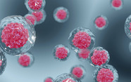
A 3D model recapitulates structure and, if done right, disease phenotype outside the body. By “done right” I mean exhibiting four qualities: biochemical markers, matching disease histopathologically (as a 3D model can be sectioned and stained), matching disease transcriptomics, and demonstrating that clinically active drugs are active, and failed clinical drugs do not work. It cannot be stressed enough that end users must have a proper standard for accepting a model – I have seen research organizations “buy in” on a model based simply on its ability to reduce the mRNA of a single disease-related marker when treated with one tool compound, and then be surprised when that doesn’t translate to success.
A well-validated 3D human tissue model is superior to animals because it offers an accurate picture of results in humans. One way to think of 3D models is that they provide ways to test the disease outside the body in a lab where extensive controlled testing can reveal the accurate response to stimuli. Every step of the process can be improved: more likely translation of validated targets; better informed structure-activity relationships and medicinal chemistry programs; better lead optimization with real data about differences between candidates prior to election for IND enabling; and better understanding of a drug’s activity during clinical trial phases, which can inform trial design.
Are the world’s regulators likely to accept a move away from in vivo testing?
I believe they are already supporting and seeking to support such a move, when the right supporting science can be presented to them. What will not change, and for now should not, is that we still need animal testing for absorption, distribution, metabolism, and excretion studies, as well as systemic tox or other issues related to the whole organism. A 3D liver model used in parallel, however, can flag liver toxicity that otherwise might be missed.
Aside from areas where animals can’t yet be replaced, regulators are science-based and have and will always accept scientific data and arguments that are convincing. As in the example of Vertex, regulators saw the value of the 3D human cell model and the data it yielded – which no animal model could provide. They now allow the use of this model to approve use of the drug in completely new patient populations – a regulatory approval for expanded use in new patients based only on in vitro results!
How long will it be before animal models are abandoned?
I’m estimating about 15 years. There is no question that 3D modelling can be superior to animal models because we know that animal models have limits. The question is whether or not the technology is ready. I think that recent results, including superior demonstration of a disease phenotype such as that published by David Brenner of Sanford-Burnham and a team at Viscient Biosciences, attesting of precision oncology applications in 3D models, better prediction of liver toxicity in complex in vitro models, and clinical phase drug programs advancing after using 3D modelling, mean that the technology is definitely worth further investment.
The first drugs that come from 3D animal tissue models will go into clinical trials in the coming years. The remaining threshold to cross is to demonstrate that the actual rate of clinical translation to efficacy is higher using these methods. Within 5–10 years, I would hope we have an emerging signal showing whether translation has improved far beyond 8 percent.


