A Sweet Revolution
Glycan analysis poses major challenges for the biopharma industry; how can new technology lighten the load?
Ken Cook |
sponsored by Thermo Fisher Scientific
About 70 percent of preclinical and clinical candidate biopharmaceuticals are glycoproteins, with carbohydrate structures attached to amino acids in the protein. These glycan groups can have a huge impact on safety and efficacy, so accurate and efficient analysis of glycans is crucial.
In this two-part series, we’ll be talking to the scientists who are applying cutting-edge analytical science to unravel the complex role of glycans in biotherapeutics.
Pick ‘n’ Mix Glycobiology
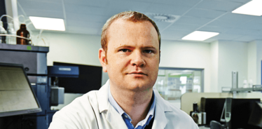
Jonathan Bones, Principal Investigator at Ireland’s National Institute for Bioprocessing Research and Training (NIBRT), uses the latest technology to help biopharma overcome challenges in glycan analysis. Here, he shares his work and divulges how using several complementary techniques is the key to (sweet) success.
What’s the mission of NIBRT?
At NIBRT, we work in close collaboration with industry partners to solve some of the key problems they face. The mix of fundamental science and real-world problems makes this a very stimulating environment; we’re applying the latest analytical science to as many aspects of bioprocessing as we can. My lab has a major focus on glycan analysis.
Why is glycan analysis so important for the biopharma industry?
It’s a regulatory requirement to provide detailed characterization of the glycan structures attached to therapeutic proteins; glycans can modify both the efficacy and safety of the molecule. For example, glycans present in the Fc region of a monoclonal antibody can modulate the interactions with Fc receptors in the immune system, which affects efficacy. In terms of safety, when proteins are expressed in non-human systems, such as CHO cells, you run the risk of non-human epitopes on glycan groups, which could elicit an immune reaction in the patient.
New technology allows us to make informed choices throughout the bioprocess, from selecting a cell line, to process testing, to the final product.
How are glycans analyzed?
A typical approach would be to detach the sugars from the protein and attach a fluorescent tag, then use high-performance liquid chromatography (HPLC)/ultra-HPLC or capillary electrophoresis (CE) to separate the fragments by size and polarity. Once you’ve separated the glycans, you usually want to characterize their structure. One method is exoglycosidase digestion, which uses enzymes to break down the sugars in a very specific and sequential manner. By looking at what you have removed and what remains, you can fit the puzzle pieces together to work out the structure. The other key technology is mass spectrometry (MS), typically used in combination with LC or CE to give you the full picture.
You make it sound relatively straightforward…
Not exactly! One of the biggest challenges is the complexity of glycans. The sequence of a protein is linear – you can visualize it as a string of beads – and we can use enzymes to break apart the beads for analysis in a predictable manner. Glycans, far from being linear, are complex branched molecules with multiple points of connection – more like LEGO® blocks than beads. That adds huge complexity because we not only have to identify the sequence of monosaccharides that makes up the glycan, but also their position and linkage orientation.
Glycan analysis for monoclonal antibodies is hard enough, but when you start looking at the larger therapeutic proteins like interferons, recombinant hormones or erythropoietins, it’s a whole new ball game, with large, complex branching glycans and modifications with inorganic substituents or sialic acids.
To unravel this complexity, you need not just one analytical technique but a range of complementary, orthogonal techniques to confirm that what you found with the first technique is what’s truly there.
How are you helping to overcome these challenges?
Right now, we’re doing exciting work on new technology for quantitative and full structural characterization of glycans in biotherapeutics. We’re starting to adopt advanced technologies from proteomics – we’re robbing the proteomics toolbox and making it our own!
Over the past five years there has been a lot of progress in quantitative analysis of glycans, with new tandem mass tags and isotope labels being developed. Currently, most analyses rely on relative results – so it’s sometimes hard to be sure whether seeing the same-sized peak on a chromatogram indicates exactly the same glycan profile. We are looking at new, stable isotope differential labeling methods, in which two independent samples are labeled separately, then run together (multiplexed) through the same LC-MS analysis. Such an approach allows us to minimize the technical variation and identify genuine changes in the molecules.
What technological advances have had big impacts on your work?
In recent years, new analytical technology has made it easier to generate the information we need. High-resolution accurate MS has really helped us to nail down structures and characterize modifications with confidence. We also do a lot of MS/MS work, using negative-ion MS to both sequence the glycan and provide additional structural and positional information. Ion mobility MS is another technology we are exploring as an add-on to LC separations – it provides another level of selectivity.
What makes glycan analysis such an exciting area?
The complexity in glycobiology gives you a lot of scope as a researcher, plus new technology and methods become available all the time. My background was in small molecules, but when an opportunity came up to work with Professor Pauline Rudd here at NIBRT, and subsequently Barry Karger at the Barnett Institute, Northeastern University in Boston, I couldn’t resist taking on a new analytical challenge.
The most satisfying aspect is seeing the science we do coming to fruition – working with the team here to translate research concepts into actual solutions that benefit the industry, and ultimately the patients.
Instrumental Sugar Rush
With Ken Cook
In the past, it’s been difficult to measure and analyze glycans – carbohydrates in general have weak polarity, do not easily stick to common column types and are difficult to detect after separation. But times have changed. More effective columns, advanced mass spectrometers, and new fluorescent reagents have made glycan analysis faster and easier. This, in turn, has generated ever-increasing interest in this field, and new advances for biopharmaceutical companies.
Safety is the top concern for all drug manufacturers. It’s crucial that anti-self glycans are not inadvertently included in glycoprotein therapies, or they could potentially kill rather than cure. Glycans also play an important role in efficacy and can act as highly effective biomarkers. For example, changes in the complex glycan structure of serum glycoproteins can be used to detect heavy drinking – something that patients often lie about.
So how can new technology help harness this potential? First, the ease of analysis has improved greatly. The first really effective columns for glycans were all amide hydrophilic interaction liquid chromatography (HILIC) columns, which can only separate by size and heterogeneity. Now, we’ve brought out two new columns that can also separate different charge states. These are particularly suitable for complex glycans, such as those found in serum proteins, which can have up to six charge states.
Monoclonal antibodies typically have much simpler glycan groups, and with the high-resolution mass spectrometers that have come out in the last couple of years, such as Thermo Scientific™ Orbitrap™-based instruments, we are now able to analyze the whole antibody at once, including any glycan groups. A single analysis obviously offers a big time-saving compared with the traditional method of deglycosylating the protein, separating out the carbohydrate, adding fluorescent labels and then carrying out liquid chromatography, often coupled with mass spectrometry (LC-MS).
Of course, getting good data from chromatography or mass spectrometry is only helpful if you can interpret it. In recent years, there has been a lot of work done on bioinformatics, both by universities and vendors, and there are now several software packages (including SimGlycan® from PREMIER Biosoft) available that can accurately identify the glycan structure from the results of an analysis.
In combination, these new techniques and technologies are allowing biopharma companies to characterize glycans with more accuracy and in more detail than ever before.
EU Bio-Separations Manager at Thermo Fisher Scientific.


